CBCT: Imaging Beyond Dental X-Rays
Radiographic examination is essential in diagnosis and treatment planning in dentistry. The interpretation of an image can be altered by the anatomy of both the teeth and surrounding
structures. The amount of information gained from conventional x rays is limited.
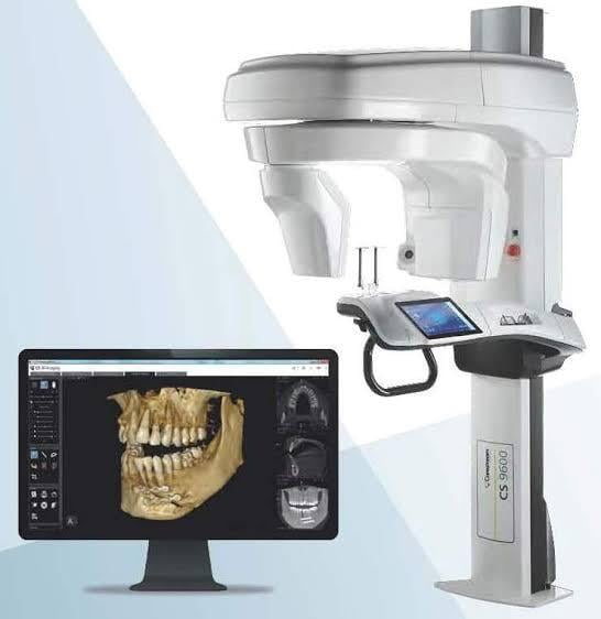
Thus came into play, the dental cone beam computed tomography (CBCT), which is a special type of x-ray machine used in situations where regular dental or facial x-rays are not sufficient. With cone beam CT, an x-ray beam in the shape of a cone is moved around the patient to produce a large number of images. CBCT produces high-quality images.
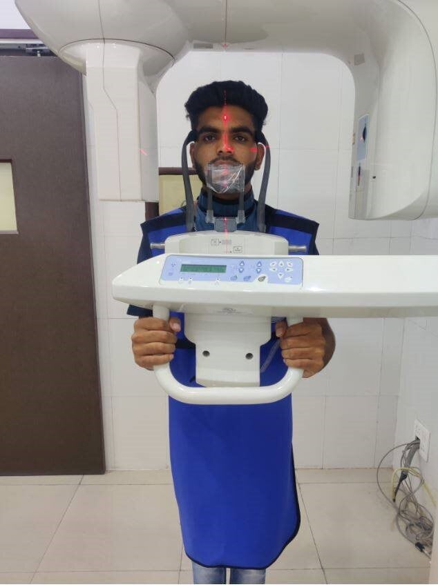
Patient Preparation
A cone beam CT examination requires no special preparation. Prior to the examination, the patient may be asked to remove metal objects, such as jewelry, eyeglasses, hairpins and hearing aids. Although removable dental work may need to be removed, it is advisable to bring these along, as the dentist may need to examine them.
Women should always inform their dentist if there is any possibility that they are pregnant.
Uses of CBCT:
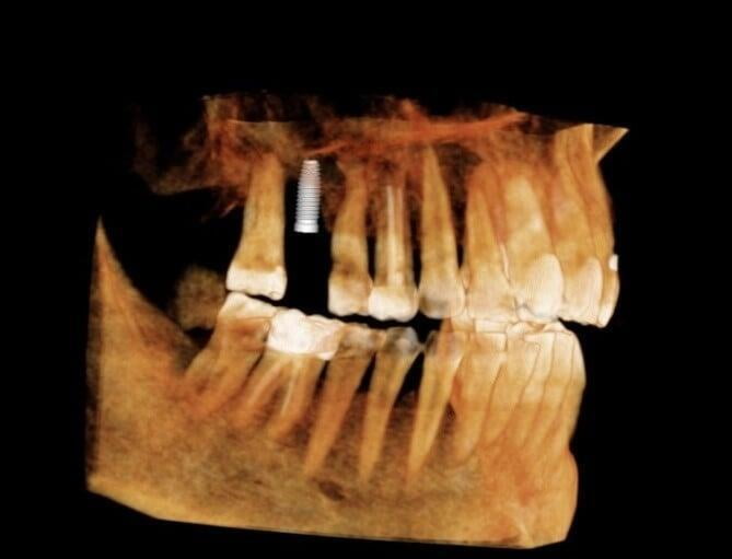
1. Implantology
CBCT can give a clear picture about the quality of bone in which the implant is to be placed, the placement in relation to the surrounding structures and in cases where bone reconstruction may be required.
2. Surgery
.
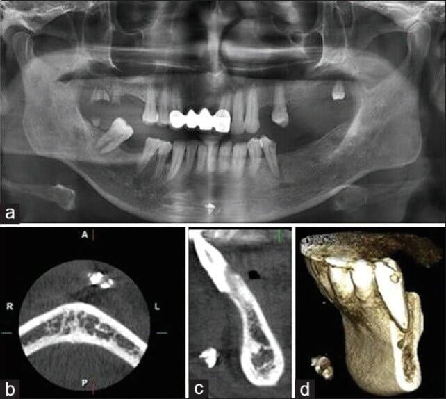
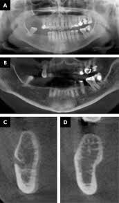
3. Trauma
CBCT is able to show a larger number of fracture lines and fractures when compared with conventional images, depicting precisely the position and orientation of displaced fragments in a reasonably short time interval.
These could be tooth and socket fractures, fractures of the orbit, fractures of the jaws or any other structure of the face and oral cavity.
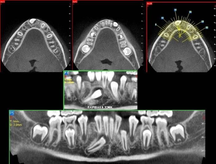
4. Orthodontics
While planning orthognathic surgeries, to evaluate the amount of airspace, to evaluate clefts in the palate, for analysis of facial structures, teeth and jaws, to place braces, to assess facial growth, age or disturbances in the eruption of teeth.
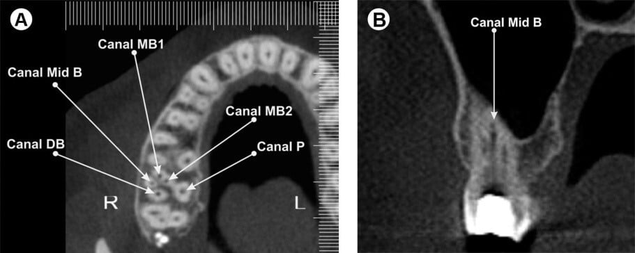
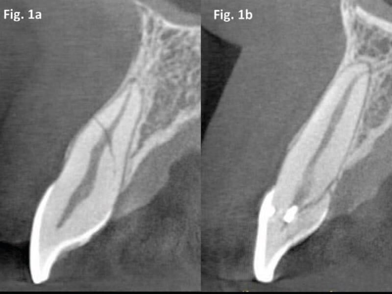
5. Endodontics
To evaluate the presence of superimposed or extra root canals, the presence of infection or pathologies around the tooth root.
6. Periodontology
CBCT allows for highly accurate analysis of bone loss as well as bone healing after periodontal treatment or regenerative therapy.
7. Forensic Dentistry
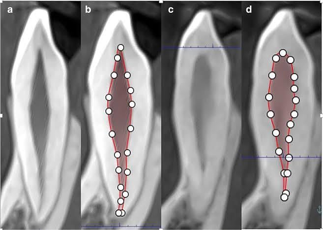
CBCT was established as a non-invasive method to estimate the age of a person based on the pulp–tooth ratio.
The team at ITS Dental College Greater Noida is highly trained and equipped in the techniques of CBCT imaging, analyzing these images and providing the best possible treatment for our patients accordingly!
Sources:
Alamri HM, Sadrameli M, Alshalhoob MA, Sadrameli M, Alshehri MA. Applications of CBCT in dental practice: a review of the literature . 2012;60(5):390-400.
Shanmugasundaram Karpagavalli. Application Of CBCT In Dentistry 2011. J Indian Acad Oral Med Radiol. 23. 163-167